Back Bones Diagram : Lower Back Muscle Anatomy And Low Back Pain : Human anatomy for muscle, reproductive, and skeleton.
Back Bones Diagram : Lower Back Muscle Anatomy And Low Back Pain : Human anatomy for muscle, reproductive, and skeleton.. The spine diagram the spine diagram shown below, consists of many bones or vertebrae,soft discs,the spinal cord, and spinal nerves. Lateral labeled diagram of the human vertebral spinal column showing vertebrae numbering order and the 5 different regions of the spine. L1, l2, l3, l4, and l5. Related posts of human back bones diagram female pelvis bones images. The first seven bones (vertebrae) of your spine form your neck.
Human anatomy for muscle, reproductive, and skeleton. For more anatomy content please follow us and visit our website: The human body is an incredible machine. Individual anatomical structures include 2: 12 photos of the human back bones diagram.
1 from At the back of each bone in the spine (vertebra) are bony points called processes, which muscles attach to. See lumbar spine anatomy diagram stock video clips. Diagram of a human female skeleton, back view. The lumbar spine connects to the thoracic spine above and the hips below. Anatomynote.com found anatomy of back muscles diagram from plenty of anatomical pictures on the internet. The vertebral column, also known as the backbone or spine, is part of the axial skeleton.the vertebral column is the defining characteristic of a vertebrate in which the notochord (a flexible rod of uniform composition) found in all chordates has been replaced by a segmented series of bone: It connects with the collarbone at the front of the body. It consists of 5 lumbar vertebra that are numbered 1 through 5 from top to bottom i.e.
The vertebral column of the lower back includes the five lumbar vertebrae, the sacrum, and the coccyx.
We think this is the most useful anatomy picture that you need. Human back bone chart, find out more about human back bone chart. The bones of the pelvis and lower back work together to support the body's weight, anchor the abdominal and hip muscles, and protect the delicate vital organs of the vertebral and abdominopelvic cavities. The lumbar spine makes up the the lower end of the spinal column. Anatomynote.com found anatomy of back muscles diagram from plenty of anatomical pictures on the internet. It also covers some common conditions and injuries that can affect the back. Spinal anatomy is a remarkable combination of strong bones, flexible ligaments and tendons, large muscles and highly sensitive nerves. The spine diagram the spine diagram shown below, consists of many bones or vertebrae,soft discs,the spinal cord, and spinal nerves. The column can be divided into five different regions, with each region characterised by a different vertebral structure. At the back of each bone in the spine (vertebra) are bony points called processes, which muscles attach to. The spine is made of 33 individual bones stacked one on top of the other. These bones are connected at the back with specialized joints. The vertebral column is a series of approximately 33 bones called vertebrae, which are separated by intervertebral discs.
It connects with the collarbone at the front of the body. But, they are common in the back and can cause pain. The anatomy of the lumbar spine is quite complex. The atlas is a ring of bone made up of two lateral masses joined at. The spine anatomy is a complex structure.
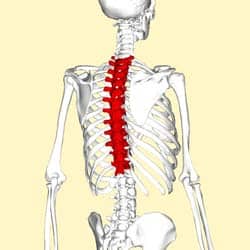
The Thoracic Spine Features Joints Ligaments Teachmeanatomy from teachmeanatomy.info This spinal column provides the main support for your body, allowing you to stand upright, bend, and twist, while protecting the spinal cord from injury. The bones of the pelvis and lower back work together to support the body's weight, anchor the abdominal and hip muscles, and protect the delicate vital organs of the vertebral and abdominopelvic cavities. For more anatomy content please follow us and visit our website: Lateral labeled diagram of the human vertebral spinal column showing vertebrae numbering order and the 5 different regions of the spine. Atlas (c1) the atlas is the first cervical vertebra and therefore abbreviated c1. The lower part of the trapezius ascends and depresses the scapula, while the transverse or middle region of the trapezius is what retracts the. Female pelvis bones images 12 photos of the female pelvis bones images female human pelvis images, female pelvic bones images, bone, female human pelvis images, female pelvic bones images The atlas is a ring of bone made up of two lateral masses joined at.
The column can be divided into five different regions, with each region characterised by a different vertebral structure.
Here we will attempt to provide a brief overview of lumbar spinal anatomy. More commonly known as the shoulder blade, the scapula is a flat triangular bone located in the upper back. We hope this picture anatomy of back muscles diagram can help you study and research. Strong muscles and bones, flexible tendons and ligaments, and sensitive nerves contribute to a healthy spine. The bones of the pelvis and lower back work together to support the body's weight, anchor the abdominal and hip muscles, and protect the delicate vital organs of the vertebral and abdominopelvic cavities. This article looks at the anatomy of the back, including bones, muscles, and nerves. Muscle or tendon injuries can occur anywhere in the body. Back of skull (occipital bone) fused vertebrae (5) (sacrum) hand bones (metacarpals) finger bones (phalanges) heel bone (calcaneus) skull (cranium) backbone 12 photos of the human back bones diagram. While many of us take the benefits of a healthy spine for granted, spinal pain is a sharp reminder of how much we depend on our back in daily life. A tough, springy disc of cartilage sits between the vertebrae of your spine. It is designed to be incredibly strong, protecting the highly sensitive nerve roots, yet highly flexible, providing for mobility on many different planes. We think this is the most useful anatomy picture that you need.
The lower part of the trapezius ascends and depresses the scapula, while the transverse or middle region of the trapezius is what retracts the. Spinal anatomy is a remarkable combination of strong bones, flexible ligaments and tendons, large muscles and highly sensitive nerves. The lumbar spine makes up the the lower end of the spinal column. Related posts of human back bones diagram female pelvis bones images. It contains the osteology, arthrology and myology of the spine and back.
Anatomy Of The Spine J J Medical Devices from www.jnjmedicaldevices.com Human back bone chart, find out more about human back bone chart. Individual anatomical structures include 2: While many of us take the benefits of a healthy spine for granted, spinal pain is a sharp reminder of how much we depend on our back in daily life. The bones of the pelvis and lower back work together to support the body's weight, anchor the abdominal and hip muscles, and protect the delicate vital organs of the vertebral and abdominopelvic cavities. From the front (or anterior), the vertebral body appears rounded. The spine diagram the spine diagram shown below, consists of many bones or vertebrae,soft discs,the spinal cord, and spinal nerves. A tough, springy disc of cartilage sits between the vertebrae of your spine. Lateral labeled diagram of the human vertebral spinal column showing vertebrae numbering order and the 5 different regions of the spine.
They help support particular bones and make them move.
More commonly known as the shoulder blade, the scapula is a flat triangular bone located in the upper back. It contains the osteology, arthrology and myology of the spine and back. The vertebral column, also known as the backbone or spine, is part of the axial skeleton.the vertebral column is the defining characteristic of a vertebrate in which the notochord (a flexible rod of uniform composition) found in all chordates has been replaced by a segmented series of bone: The bones of the chest and upper back combine to form the strong, protective rib cage around the vital thoracic organs such as the heart and lungs. This spinal column provides the main support for your body, allowing you to stand upright, bend, and twist, while protecting the spinal cord from injury. Here we will attempt to provide a brief overview of lumbar spinal anatomy. Spinal anatomy is a remarkable combination of strong bones, flexible ligaments and tendons, large muscles and highly sensitive nerves. The spine is made of 33 individual bones stacked one on top of the other. Human back bone chart, find out more about human back bone chart. Your lower back contains 5 vertebral bones stacked above each other with intervertebral discs in between. The spine diagram the spine diagram shown below, consists of many bones or vertebrae,soft discs,the spinal cord, and spinal nerves. The first seven bones (vertebrae) of your spine form your neck. At the back of each bone in the spine (vertebra) are bony points called processes, which muscles attach to.
0 Response to "Back Bones Diagram : Lower Back Muscle Anatomy And Low Back Pain : Human anatomy for muscle, reproductive, and skeleton."


0 Response to "Back Bones Diagram : Lower Back Muscle Anatomy And Low Back Pain : Human anatomy for muscle, reproductive, and skeleton."
Post a Comment